Often the first exam during an audiology appointment, otoscopy is a non-invasive medical procedure used to examine the external ear canal and the eardrum (tympanic membrane). It involves the use of an otoscope, a handheld device equipped with a light source and magnifying lens. This tool allows hearing healthcare professionals to visualize the structures of the ear and identify any abnormalities.
AUDIOLOGY PRACTICE MANAGEMENT SOFTWARE

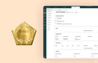
Auditdata Manage, a cloud-based, data-driven practice management software, optimizes audiology practices with paperless protocols, automated tasks, and insightful patient management, enabling higher quality care and efficiency.
Learn more about ManagePRACTICE MANAGEMENT SOFTWARE
-
Explore all our features
Discover the extraordinary features of our Practice Management Software. -
Reporting
Easily access reports of sales, operations, finance, marketing, and administration. -
Integrations
Connect, streamline, and improve audiology workflows with integrations. -
Onboarding and Support
Get the most out of our software with top-notch onboarding and support. -
Security and Configuration
Easy to configure and offer superior security features.
Audiological Equipment & Software

Auditdata Measure offers a modular, PC-based audiological testing and fitting suite that improves accuracy, patient comfort, and workflow efficiency, reducing return rates and enhancing customer satisfaction.
Learn more about MeasureComprehensive Solutions for Every Need
-
Hearing Assessment
Our comprehensive suite of hardware and software solutions designed to revolutionize the field of hearing assessment. -
Hearing Instrument Fitting
If precision in instrument fitting and thorough testing is what you seek, our integrated solution is designed for you. -
Reporting
Tap into powerful analytics and performance metrics, our clinical and fleet reporting tools offer insights into your practice's health. -
Services
Our transducer swap service and comprehensive training programs are perfect solutions for you.
Looking for Equipment and Software?
-
NEW
Audiometer & Fitting Unit
The new Measure audiometer and fitting unit solution offers flexibility through wired or wireless accessories. -
Tympanometer
A battery-operated, portable diagnostic tympanometer that offers unparalleled flexibility and top-of-the-line features. -
HIT box
Explore the Hearing Instrument Tests box and learn how to verify the technical specifications of a hearing aid. -
Updated
Measure Software
Utilize our Measure Software, formerly known as Primus, to elevate clinical efficiency and enrich patient care.
iPad Hearing Screener

Auditdata Engage transforms early hearing impairment screening with an iPad-based solution that engages potential customers, streamlines lead conversion for audiologists and clinics through data-driven pre-screening insights.
Learn more about EngageMost Popular Features
-
Customize Screener Flow
Customizable modular elements to help you build your own test flow for any setting or location. -
Configure Testing Protocols
Configure determination, designating corresponding values for some or significant hearing loss ranges. -
Add Clinic Branding
Enhance your clinic's brand identity with a fully customized, visually aligned hearing screener. -
iPad Screener Reports
Track screenings, prioritize them, and nurture each lead to help convert prospects to patients.
Tailored for Small and Medium-Sized Businesses and Large Enterprises
-
Engage | Hearing Screener
Self-service screening and lead capture. Build your pipeline in the most cost-effective way. -
Measure | Audiological Equipment & Software
Portfolio of audiological equipment run by advanced software. Take your clinical performance to the next level. -
Manage | Practice Management Software
Data-driven cloud-based practice/office management software. Everything you need to run and grow your audiology business. -
Discover | Add-on BI tool
Analytics and performance benchmarking tool. Turn your data into competitive advantage.
Hospital Solutions
View all-
Tailored for public audiology
We build solutions specifically tailored to the needs of a fast-moving public audiology clinic -
Patient flow optimisation
Manage your patients better, optimise your workflows and reduce your waiting lists -
Optimise your clinic
Run a successful audiology clinic with our built-in data analysis and start optimising your clinic -
Facilitating ENT, CI and Otology Surgeons
ENT Doctors, Cochlear Implant and Otology surgeons can communicate with their Audiology teams, reducing wait times and costly miscommunications.
Get a Demo
Schedule a demo today!
See how Auditdata can help you transform the way your practice will attract and retain customers by efficiently manage and deliver your services with increased sales and happier customers.

Best Care Experience
-
Pre-clinic: Awareness and screening
Explore ways to close the gap from detection to treatment with tools for awareness, self-screening, and pre-qualifications of patients. -
In-clinic: Testing and fitting
Discover ways to enhance testing and fitting accuracy, enabling more meaningful professional-patient interactions. -
Aftercare: Adoption and Follow-up
Learn how structured follow-ups and reminders reduce hearing aid return rates and support successful adoption.
Insights
GO TO ALL INSIGHTS-
Blog
Find the latest articles on the hearing care industry and products. -
Ebooks & Guides
Guides on relevant topics in the hearing care industry -
Customer stories
Discover how Auditdata is helping clinical business' grow without compromising patient care -
New
Personal Stories
Explore unheard personal stories of people living with hearing impairment.
Auditdata Webinars
Explore Our Latest Webinars
Stay ahead in audiology and explore our library of engaging webinars where industry experts dive into the latest trends, tools, and strategies to help you provide best care experiences to all your patients.
Go to WebinarsCompany
-
About us
Learn about Auditdata and discover what drives us -
Meet Our Global Team
With over 30 nationalities represented, our team of 150+ dedicated professionals spans the globe. -
Management
Meet our Management Team -
Board of Directors
Meet our Board of Directors -
Careers
We are always looking for talented, dedicated people -
News
Get the latest news and industry insights right here. -
Download Annual Report 2022
Learn more about our financial, social, and environmental performance in one integrated report. -
Environment Social Governance
We drive positive impact and sustainable growth across our organization and beyond.
Get in touch
Talk to a member of our team
We’ll help you find the right products and pricing for your business

Explore our Help Center
A knowledge base of how-to-guides, information, documentation and FAQs for Auditdata's products

Shortcuts
-
Create Support Ticket
If you have any questions or feedback, feel free to submit a ticket and our support will help you -
Calibration service
With Auditdata SWAP service we ship calibrated transducers to you well in advance of the due date of the old ones. -
Contact support
We offer comprehensive support and training to all our customers. Read our Service Level Agreement. -
Terms and conditions
The following terms and conditions apply to all services and deliverables provided by Auditdata A/S

